About Me

Computed tomography, CT
• linear tomography
• principles of CT
• equipment
Linear, conventional tomography
• Synchronized movement of x-ray tube and
film about the fulcrum or axis
• Longitudinal sections
• Axis = tomographic plane
• Thickness of the plane:
5 6
Linear, conventional tomography
• Tube movements
– Linear
– Non linear
• circle
• ellipse
• lissajous
• spiral
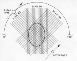
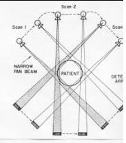
Principle of CT
• x-ray source rotating around the patient
• Detector array on the opposite site of the
patient
• Up to 1000 projections are measured in
angles covering 360o
• Object is represented by an matrix of volume
elements (voxels) shoving individual
attenuation coefficient m
• Beam attenuation is measured =
ray sum

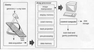
CT equipment
• CT-systems
Tomography
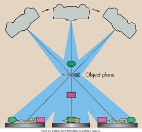
Only objects lying in the object plane are properly imaged. Objects above and below this plane are blurred because they are imaged across the film.
Fluoroscopy

What Is Flouroscopy?
Fluoroscopy is the study of moving internal body parts. A continuous x-ray beam is passed through the body part being examined, and is transmitted to a TV-like monitor so that the body part and its motion can be seen in detail. During Fluoroscopy the x-ray tube is operated at less than 5 mA.
2
Fluoroscopy, as an imaging tool, enables physicians to look at many body systems, including the skeletal, digestive, urinary, respiratory, and reproductive systems. Fluoroscopy may be performed to evaluate specific areas of the body, including the bones, muscles, and joints, as well as solid organs such as the heart, lung, or kidneys
3
Fluoroscopy is used in many types of examinations and procedures, such as barium x-rays, cardiac catheterization, arthrography (visualization of a joint or joints), lumbar puncture, placement of intravenous (IV) catheters (hollow tubes inserted into veins or arteries), intravenous pyelogram, hysterosalpingogram, and biopsies.
4
Fluoroscopy may be used alone as a diagnostic procedure, or may be used in conjunction with other diagnostic or therapeutic media or procedures.
In barium x-rays, fluoroscopy used alone allows the physician to see the movement of the intestines as the barium moves through them. In cardiac catheterization, fluoroscopy is added to enable the physician to see the flow of blood through the coronary arteries in order to evaluate the presence of arterial blockages. For intravenous catheter insertion, fluoroscopy assists the physician in guiding the catheter into a specific location inside the body
In barium x-rays, fluoroscopy used alone allows the physician to see the movement of the intestines as the barium moves through them. In cardiac catheterization, fluoroscopy is added to enable the physician to see the flow of blood through the coronary arteries in order to evaluate the presence of arterial blockages. For intravenous catheter insertion, fluoroscopy assists the physician in guiding the catheter into a specific location inside the body

ABC
(ABC) stands for Automatic Brightness Control, and it is mostly controlled by changing the Kvp and mA. This is done to get the same brightness Image. But The risk in increasing this is increasing the patient dose.
PP
PP2
PP3
PP4
PP5
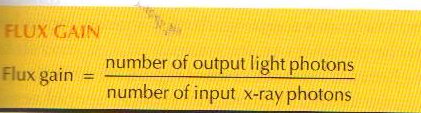
Flux Gain
Brightness Gain

Minification Gain
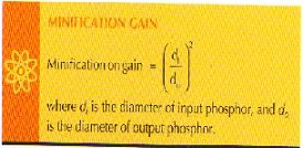
The minification gain is the ratio of the square of the diameter of the input phosphor to the square of the diameter of the out put phosphor.
Conversion Factor
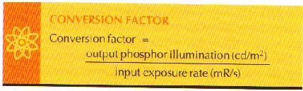
(cd/m*) = candela per meter squared.
(mR/s) = milliroentgens per second.
(mR/s) = milliroentgens per second.
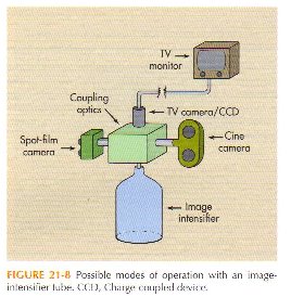
CCD


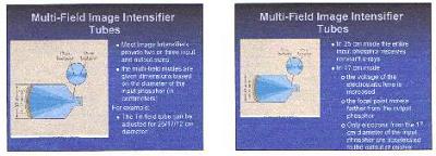
When one switches from the 25 cm mode to the 17 cm mode in a multi-field tube:
- The input field of view is reduced.
- The image appears magnified on the output screen.
- The Magnification Fator becomes 1.5.
- The Image gets dimer.
-The mA is increased to compensate Image Brightness.
- The input field of view is reduced.
- The image appears magnified on the output screen.
- The Magnification Fator becomes 1.5.
- The Image gets dimer.
-The mA is increased to compensate Image Brightness.
Digital Fluoroscopy
Digital fluoroscopy is a form of x-ray that allows us to view deep structures of the body in real time. It provides very detailed images of function and structure of areas like the intestines, the bladder, the cardiac muscle and stomach. Unlike regular x-ray which records the image to film, digital fluoroscopy records a series of images to a computer. Once digitized, we can view the area being examined in real time on a computer monitor.
How Digital Fluoroscopy Works
Digital fluoroscopy uses a controlled beam of energy that is passed through the body and captured by an image detector. Because the bones, organs and tissues within our bodies are composed of differing densities, the beams move through them differently. Bones for instance will absorb more of the beam than an organ or soft tissue making them appear white or gray on the image while the tissue appears darker.
The image information passes through a one-million pixel camera where it is digitized and sent to the computer. The conversion is immediate and yields a high resolution digital image that we can view much like a movie of what is happening inside the body.
Sometimes digital fluoroscopy makes use of a contrast agent like barium. These agents are radio-opaque liquids which provide a white appearance on the fluoroscopic image. As the agent moves through the exam area, generally an organ, the radiologist is able to track its path and evaluate the organ as it functions as well as size and location.
CCD
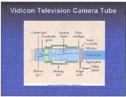
The Image Above shows a typical Vidicon. The glass enveloped serves the same function that it does for the x-ray tube. These internal elements include the cathode, its electro gun, assorted electrostatic grids, and a target assembly that serves as the anode.

The Heart of the Television Monitor

Spot Film
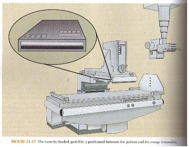
The photo spot camera uses film sizes of 70 and 105mm. As a general rule, larger film formats results in better image quality but increases patient dose.
TV Monitor
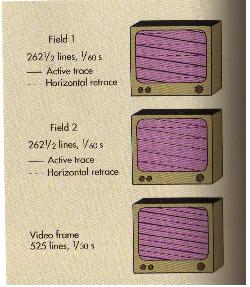
TV monitor uses a rate of 30 frames per second.
Thursday, January 22, 2009
Subscribe to:
Posts (Atom)